Oral Surgery
What Is a Dental Bone Graft? A Complete Patient Guide
A dental bone graft rebuilds jawbone where a tooth was lost. What it is, when it's needed, why bone disappears, and how it restores volume for an implant.
Written by Dr. Husna Khan, DDS
Serenity Dental of Bloomingdale · April 29, 2026
Educational purposes only. Whether a graft is needed in your case depends on CBCT measurements taken at consultation. Call (630) 359-0105.
A dental bone graft sounds intimidating. The word “graft” pulls up images of major orthopedic surgery. In dentistry, it is something far smaller and far more routine — a thirty-to-ninety-minute outpatient procedure that adds a small volume of bone material to a thin spot in the jaw, so a future dental implant has a stable foundation.
This guide explains exactly what a bone graft is, why jawbone disappears after a tooth is lost, when grafting is genuinely needed, and what the recovery looks like.
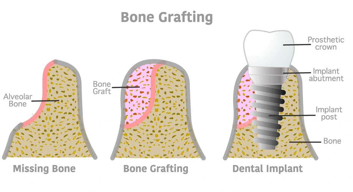
A dental bone graft, defined
A dental bone graft is a small piece of bone or bone-substitute material that is placed into a deficient area of the upper or lower jaw. Once placed, it acts as a biological scaffold. Over 3 to 6 months the patient’s own cells migrate into the scaffold, lay down new living bone tissue, and gradually replace the graft material with native bone. The Cleveland Clinic describes the same biological process: the graft is not a permanent foreign object but a template that the body uses to grow its own bone.
A typical graft is a few millimeters of granular material — about the size of a pencil eraser, sometimes smaller. It is placed in a controlled surgical site under local anesthesia. A thin barrier membrane is then placed over the surface to keep gum tissue from growing into the area before bone has had time to form. Sutures close the gum on top.
That is the entire procedure. It is closer in scale to a tooth extraction than to any kind of orthopedic surgery.
Why does jawbone shrink when a tooth is lost?
Ridge width over time after tooth loss

Jawbone is unusual among the bones in the body. It does not maintain its volume on its own. It needs the daily mechanical pressure of chewing — transmitted through the tooth root into the bone — to stay thick and dense.
When a tooth is lost, that pressure signal stops. The body interprets the bone in that area as no longer needed and gradually resorbs it. The American Academy of Periodontology reports that the alveolar ridge can lose up to 25 percent of its width within the first year after a tooth is removed. Most of that loss happens in the first 3 to 6 months. After year one, the rate slows but loss continues.
This is why the timing of an extraction visit matters so much. A graft placed into the empty socket immediately after extraction — called socket preservation — keeps the ridge from shrinking in the first place. Waiting six months and then trying to rebuild the lost volume is a larger procedure than preventing the loss to begin with.
When do you actually need a bone graft?
Not every implant patient needs a graft. That decision is made from a 3D cone-beam CT scan that measures bone height, width, and density at the exact site where the implant would go. A CBCT scan is non-negotiable in implant planning — without it, the dentist is guessing.
A graft is needed when the scan shows insufficient volume for the implant length and diameter the case requires. Common scenarios that produce that finding:
- Tooth lost more than a year ago without replacement. The longer the gap, the more bone has resorbed.
- Tooth lost to periodontal disease. Gum disease destroys the bone around the tooth before the tooth itself comes out, so the starting volume is already low.
- Upper back-jaw implants near the sinus. The maxillary sinus naturally hangs down close to where molar roots used to anchor. A sinus lift adds bone height between the jaw and the sinus floor.
- Long-term denture wearers. Removable dentures do not transmit chewing pressure into the bone the way teeth do. After a decade the ridge often becomes very thin.
- Trauma or infection that damaged the bone before the tooth came out.
A graft is not needed when the scan shows adequate height and width and good bone density — most commonly seen in patients who had the tooth removed recently and have otherwise healthy bone.
In our office, when CBCT shows borderline volume, Dr. Khan will always show the patient the actual measurements rather than just stating a recommendation. Seeing the numbers — millimeters of available bone versus millimeters needed for the planned implant — usually answers the question on its own.
What does the procedure involve?
Bone graft procedure — five steps

A bone graft for a single site typically follows the same five-step pattern. The whole appointment usually runs 30 to 90 minutes depending on how much bone needs to be added and whether grafting is being combined with an extraction or implant placement.
Local anesthesia numbs the patient first. Optional IV or oral sedation is available for anxious patients but is not medically required for the procedure itself. The gum is opened with a small incision to expose the bone surface. Graft material is packed into the deficient area. A barrier membrane is laid over the graft to protect it from gum tissue ingrowth. The gum is then sutured closed over the membrane.
Recovery follows a familiar pattern for any oral surgery. Mild swelling and soreness peak around days 2 to 3 and ease over the first week. Most patients describe the experience afterward as similar to a routine tooth extraction — easier than they expected. The graft itself works invisibly underneath. Externally, the site looks normal while bone forms underneath over the next 3 to 6 months.
What is the graft made of?
Four materials are used in modern dentistry. Each has a long clinical history and well-documented success rates.
Allograft — processed bone from a human donor. The most common choice for routine dental implants. Tissue is fully sterilized and screened. There is no second surgical site on the patient. The American Association of Oral and Maxillofacial Surgeons recognizes allograft as a standard, safe option.
Xenograft — processed bovine bone, most often. The mineral structure of cow bone is similar to human bone, which makes it an excellent scaffold. Bio-Oss is the best-known brand.
Alloplast — fully synthetic graft material, typically calcium-based ceramics like beta-tricalcium phosphate. No biological tissue at all. A common choice for patients who prefer a non-animal, non-donor source.
Autograft — the patient’s own bone, harvested from another site such as the chin or behind the wisdom tooth area. Used in larger reconstructions but rarely needed for single-implant cases because of the second surgical site.
For most single-tooth cases at our office, Dr. Khan uses allograft or a synthetic alloplast. Both eliminate the need for a second surgery and have decades of evidence supporting predictable results.
Healing — what is happening invisibly
Once placed, the graft begins a biological process called osseoconduction. The graft material is porous. The patient’s own bone-forming cells, called osteoblasts, migrate into the pores from the surrounding native bone. Those cells lay down new collagen matrix, mineralize it, and gradually replace the graft particles with living bone.
This timeline is consistent across patients. New blood vessels form within the first 2 weeks. Early bone formation is visible on imaging by 3 months. The site reaches enough density to support an implant somewhere between months 3 and 6 depending on the size of the graft and the patient’s healing biology. Larger ridge augmentations and sinus lifts are typically held to the full 6 months.
Smoking, uncontrolled diabetes, certain bone-density medications, and aggressive disturbance of the surgical site all slow this process. The single largest variable in patient-controlled outcomes is whether the patient avoids smoking during healing.
A note on cost and insurance
Single-site bone grafts at extraction (socket preservation) are typically varies in the Bloomingdale area. Larger ridge augmentations and sinus lifts run $300–$1,200 or more. Dental insurance coverage varies widely — some plans include grafting under major restorative benefits when documented as medically necessary for implant placement, others exclude it. A detailed cost breakdown is in the related guide below.
Bottom line
A dental bone graft is a small, well-established outpatient procedure that rebuilds jawbone where it has been lost. It is needed when the bone is not thick or tall enough to safely anchor a planned dental implant. The procedure takes well under two hours, recovery feels like a tooth extraction, and the site needs 3 to 6 months to mature before the implant goes in. None of it is exotic or experimental — bone grafting has been performed routinely in dentistry for decades.
If you have been told you may need a graft and want a clear, honest assessment with the actual CBCT measurements, call Serenity Dental in Bloomingdale at (630) 359-0105. Related: bone grafting service page · types of bone graft material.
Common questions about dental bone grafts
What is a dental bone graft in simple terms?
Are bone grafts always necessary?
Why does jawbone disappear after a tooth is lost?
Can I get a dental implant without a bone graft?
Is a dental bone graft considered surgery?
How long does a dental bone graft take to heal?
What happens if I do not get a recommended bone graft?
Educational content only. Recommendations are personalized after an exam and any needed imaging.
About this article
Educational purposes only and not a substitute for individualized clinical evaluation. Bone-loss timelines reflect American Academy of Periodontology guidance. Graft material categories reflect AAOMS classification of bone graft sources. Whether grafting is required in any individual case is determined by CBCT imaging at consultation.
.
Pricing note. Prices shown are approximate Chicago-area 2026 ranges compiled from public cost guides — not a quote or a Serenity Dental fee schedule. Your actual cost depends on your clinical needs, the materials chosen, and your insurance. We provide a written estimate before any treatment begins. Call (630) 359-0105 to confirm pricing for your situation.
Need help with this in real life?
Reading helps. Talking to someone who can look at your actual teeth and symptoms helps more. If you want a clear next step, we’re here.
Related articles
Types of Dental Bone Grafts: Autograft, Allograft, Xenograft, and Synthetic
The four bone-graft materials in dentistry: what each is, where it's from, and how to choose, with guidance for religious, ethical, or vegan concerns.
Read article →Broken Tooth Extraction: What to Expect
How broken tooth extraction differs from simple extraction -- surgical approach, cost, recovery, and whether a regular dentist can remove a broken tooth.
Read article →Infected Tooth Extraction: When It's Needed and What to Expect
When an infected tooth needs extraction vs saving, antibiotics before extraction, post-op infection signs, and what recovery looks like.
Read article →